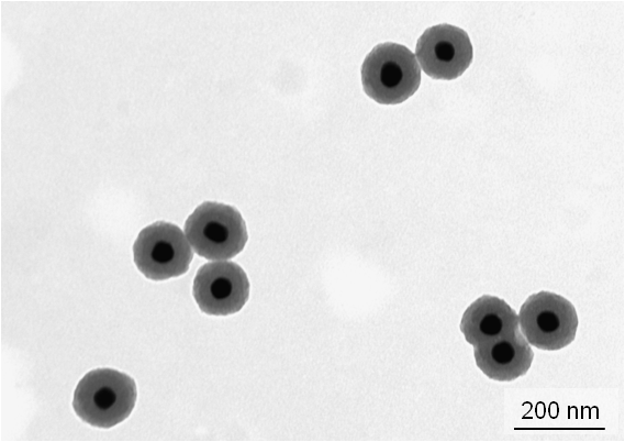
The QD/SiO 2/SH/Au/PEG nanoparticle colloidal solution was injected into the tail vein of a mouse and circulated in the blood, although some QD/SiO 2/SH/Au/PEG nanoparticles were trapped and remained in the liver and spleen. The CT value per unit concentration of Au (M) for the QD/SiO 2/SH/Au/PEG nanoparticle colloidal solution was 1.5 × 10 4 HU/M. Hereby, we present a synthetic control strategy for the large-scale production of goldsilica coreshell NPs and provide an opportunity to synthesize other NPs with different shapes and structures by PLAL. Monodisperse silicapolyaniline coreshell nanoparticles with an average diameter of 26 nm were synthesized by in-situ polymerisation of aniline monomers.

The QD/SiO 2/SH/Au/PEG nanoparticle colloidal solution was imaged by fluorescence and x-ray CT imaging techniques. PLAL provides a simple way for a one-step synthesis of coreshell NPs as has been shown by previous work. Surface-PEGylated QD/SiO 2/SH/Au particles were fabricated using thiol-terminated polyethylene glycol (PEG) (QD/SiO 2/SH/Au/PEG). The core-shell structure silica-poly(methyl methacrylate), SiO2-PMMA, nanoparticles were formed by grafting polymerization of MMA on the surface of the. Immobilization of the Au nanoparticles on the QD/SiO 2/SH particle surface was performed with the addition of the Au nanoparticle colloidal solution to the QD/SiO 2/SH nanoparticle colloidal solution (QD/SiO 2/SH/Au). (3-Mercaptopropyl)trimethoxysilane thiolated the QD/SiO 2 particle surface (QD/SiO 2/SH). Nanoparticles Used in Research Area of Strong Scientific Interest Due to The Variety of Application in Biomedical Electronic and Optical Fields Nickel/ Silica. Reduction of Au 3+ in a citrate aqueous solution gave an Au nanoparticle colloidal solution. The QD nanoparticles were coated with silica shells by a sol-gel method using tetraethyl orthosilicate (QD/SiO 2). consists of the nucleic acid and an outer shell of protein. This work describes the fabrication of quantum dot (QD)/silica (SiO 2) core–shell particles immobilized with Au nanoparticles and the evaluation of their abilities in fluorescence and x-ray computed tomography (CT) imaging. Each virion consists of a core of nucleic acid (viral chromosome or genome) and a proteinous.


 0 kommentar(er)
